An NYBG Internship Experience: Connecting the Dots to Understand Relationships between Living and Extinct Cycads
Posted in Applied Science on May 15, 2018 by Cynthia Huyck
Cynthia Huyck is a former intern at NYBG’s Pfizer Plant Research Laboratory. She is completing her undergraduate degree at Sarah Lawrence College, concentrating in botany and Japanese.
Editor’s Note: As the end of the academic year approaches, The New York Botanical Garden’s Plant Science and Conservation program will soon welcome a new class of summer interns. As described in this post, these internships offer valuable experience by allowing students to work closely with NYBG scientists on cutting-edge research.
I spent last summer and fall as an intern working with Vice President and Cullman Curator Dennis Stevenson, Ph.D., on the Gymnosperm Seed Evolution Project at The New York Botanical Garden. The goal of this project is to better understand the relationships among different gymnosperms based on their seeds and seed-bearing organs. My work focused on the anatomy of early phases of ovule development, with an emphasis on branching of the vascular bundles, which contain the specialized cells that transport water and nutrients throughout the plant. I used an anatomical approach because my work involved comparing extant and extinct cycads. Although we cannot extract DNA from the fossils, we can look for structural features in both the fossils and extant species in order to place our fossils on a phylogeny (evolutionary tree) and establish a minimum age for cycads.
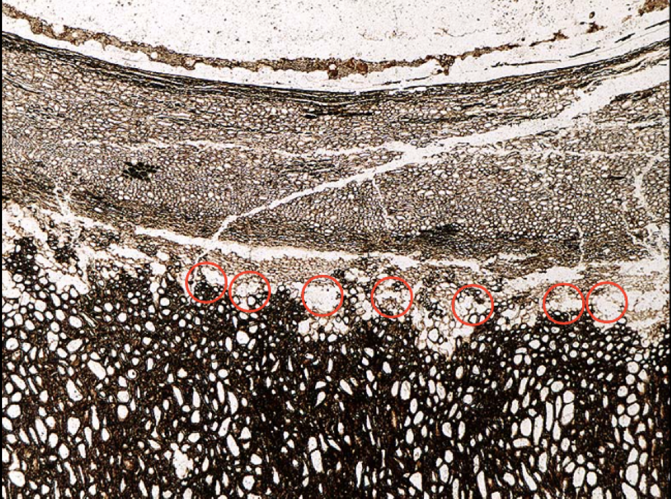
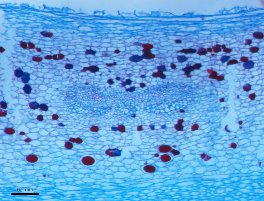
To gather the necessary data on early vascular branching in the seed coat of extant species of cycads, I used modern histological techniques to fix, section, and stain young ovules. To establish relationships, I examined the second layer of the integument (or seed coat), to determine whether the vascular bundles branch polychotomously (one branch turning into three or more branches) or dichotomously (one branch turning into two). This required sectioning ovules from the base up, in order to develop a three-dimensional idea of how the bundles connect and divide as they travel up the ovule. I found that some major genera of cycads indeed have polychotomous vascular branching. I then compared my findings to the fossil peels of extinct cycad species and found that the fossil peels show the same polychotomous vascular branching. This will help us to understand relationships between living and extinct species of cycads in order to better understand how and when they evolved.
My internship afforded me a unique opportunity to engage with science and plants in one of the nicest labs in the United States, improving my individual research skills and teamwork skills. I worked with one other intern on the project, which forced me to keep my data organized and understandable.
