Forming our Attention: An Artist turned Scientific Illustrator
Owen Roberts is an NYBG/SciNetwork NYC Intern, and a researcher on the project Podostemum ceratophyllum led by Assistant Curators Dr. Ana María Bedoya and Dr. Cecilia Zumajo, New York Botanical Garden.
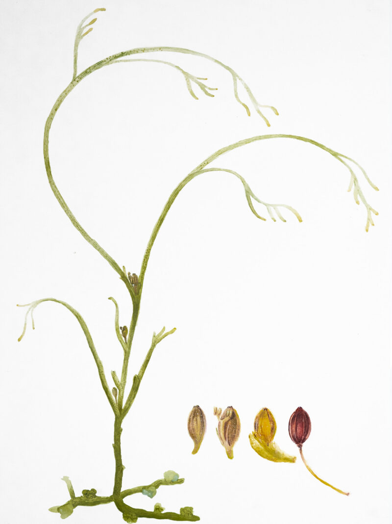
Figure 1. Watercolor of Podostemum ceratophyllum showing the vegetative plant with flowers, located at bottom right. From Left to Right: Floral bud with plethora still attached, Flower in anthesis, Flower with plethora (at base of flower) naturally falling off, and Fruit.
When I was about 10 years old, I started drawing insects from observation. I would lift up the tetherball poll and tire in my backyard, and crickets, centipedes, and pillbugs would scatter all around, some which I would catch. Then, sitting on the grass, I’d grab my blue lined notebook, peek through my fingertips to my dirty palm, which they squirmed around, and sketch them with my right hand. I didn’t know it then—it was all just play to me—but I was making scientific illustrations. Although I have always drawn animals, I discovered the world of botany and its art just this last year after stumbling upon the beautiful Hilma Af Klint botanical art exhibition at the MoMa. I knew, then and there, that I wanted to learn more about botanical studies and expand my art practice.
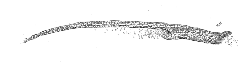
Figure 2. Illustration of the longitudinal section of a leaf.
I looked into environments which would foster this interest, and the New York Botanical Garden fit just right. I reached out hoping to learn more about opportunities and find ways to integrate scientific observations with my detailed artistic eye, and suddenly I ended up working under two botanists and curators at NYBG, Dr. Ana María Bedoya and Dr. Cecilia Zumajo. They gave me the opportunity to join their ongoing research project on Podostemum ceratophyllum, a flowering river plant in the family Podostemaceae (commonly called riverweeds). It is an odd species, not only in its overall appearance, but also in its development, anatomy, and habitat, which we know relatively little about. For example, Podostemum ceratophyllum does not have roots, stems, or leaves that can be readily distinguished from each other. Rather, botanists refer to the body as a ”thallus,” like in algae. What we are venturing to understand is where the meristematic tissue may be, and for that we need more observations. The Podostemaceae lack a clearly identified primary root system as seen in figure 1, and it attaches to rocks putatively, via a biofilm produced by cyanobacteria.
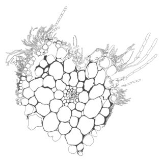
Figure 3. Illustration of the cross section of the leaf.
To understand its anatomy, we embedded the tissue into resin, which we spliced in 3um (3 micrometers) thick sheets with a microtome. Once all is cut, we placed the sheets onto glass slides, staining them with toluidine blue (a metachromatic dye which reacts to lignified tissues, turning them greenish blue, and soft parenchyma tissues, turning them pink). Once recorded, the composite images were pieced together by stacking several photographs of the cellular structures, which appear to be porous and delicate (figures 2 & 3). Observing its fruits and flowers further confirms this intricate porosity throughout the plant body (figure 4).
Many of these open questions only deepen my fascination with what is and what may be genuinely an anomalous lineage. Its thallus-like body shifts between olive green, bright green, and sometimes reddish ochre, becoming lighter on fresher material. To me, it resembles moss or algae, seemingly slimy and slippery, but they strongly attach to rocks, needing to be pried off with a knife.
Its strength is not incidental. Although the fast-moving river rapids of eastern North America subject organisms to powerful hydraulic forces and debris collision, Podostemum ceratophyllum (and the Podostemaceae in general) has adapted to thrive in one of the most mechanically demanding freshwater environments. If it were me holding onto that rock, I’d probably lose my grip and drift lazily down the river with sketchpad in hand.
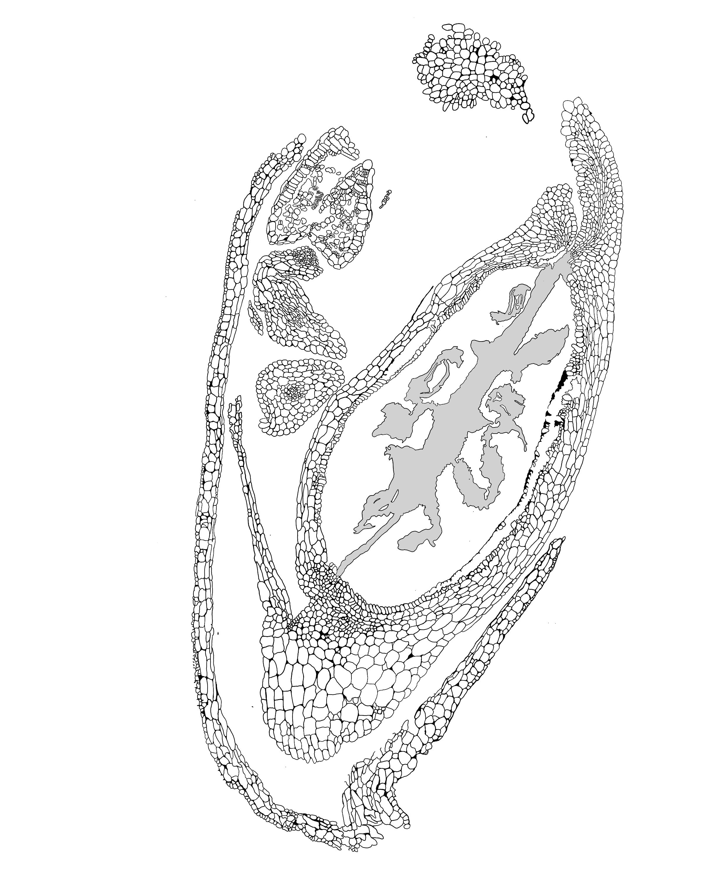
Figure 4. Illustration of the longitudinal section of the flower.
Before working in a lab, I had never illustrated internal anatomy, so I didn’t fully understand the external and internal structures of plants. Through my experience I have realized the importance of accuracy of scientific documentation. Scientific illustration must accurately depict an object, bridging beauty, clarity, and accuracy. If the illustration is accurate and not beautiful, it would join the pot of illustrations that the world already has. Yet, if an illustration was beautiful while inaccurate, it would not be useful for science and hold only the non-scientific community’s eyes. As an artist, “beauty” becomes a complicated subject which I continually navigate. However, since beginning my internship at NYBG I have started to look at plants differently.
I have always loved the jitter of tree branches in soft wind, or the cast shadows of light below a tree line, but never had I been so engaged with the way which plants grow until studying plant anatomy. My eyes, now, nearly graze the surface of their leaves and flowers, noticing their venation, ridges, and thorns. The question of where, exactly, a plant’s meristematic tissue lies has become an essential curiosity quest for me since starting this job. I’ve begun to feel the same sense of wonder I did when I lifted that tire at 10 years old, yet this time around, the specimens aren’t wiggling around in my hand.
Perhaps, that was the whole point—science, like art, is just another name for a kind of looking that we forget when we grow up.
SUBSCRIBE
Enter your email address to subscribe to this blog and receive updates on new posts.


